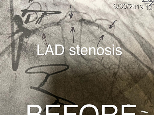
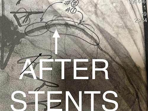
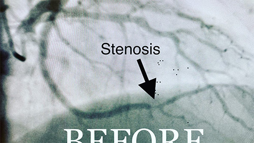
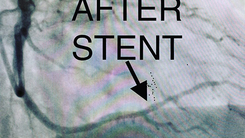
68 y/o male with diabetes, CABG(coronary artery bypass surgery) one year ago developed chest pain and small troponin leak. Coronary angiogram showed high-grade long lesion in the native LAD (bypass conduit to LAD was occluded). See BEFORE picture. The patient received two DES stents with complete resolution of the blockage (see AFTER picture). His chest pain resolved after the stent procedure.
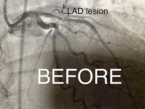
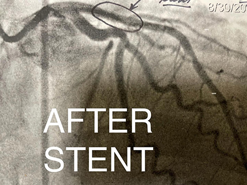
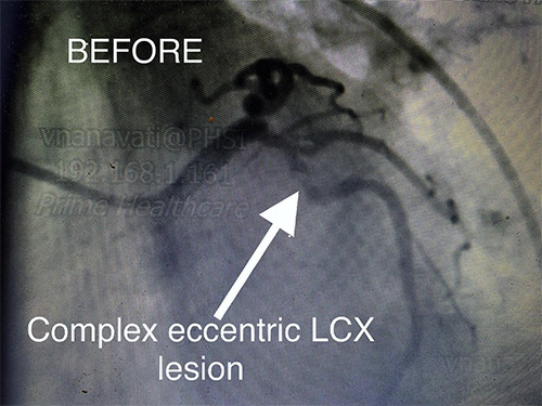
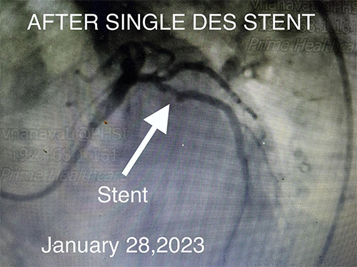
47y/o diabetic female with chest pain and dysrhythmia showing NSVT and slight elevation in Troponin. Coronary angiogram shows a severe eccentric 95% stenosis ( see BEFORE). She received a single DES stent in LAD with resultant resolution of the stenosis and excellent flow( see AFTER STENT). Her symptoms resolved after flow in the LAD was restored.
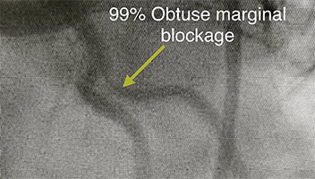
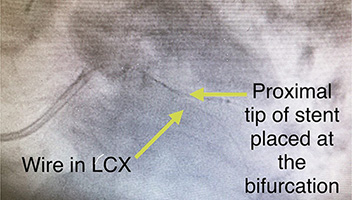
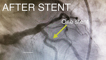
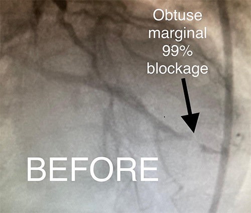
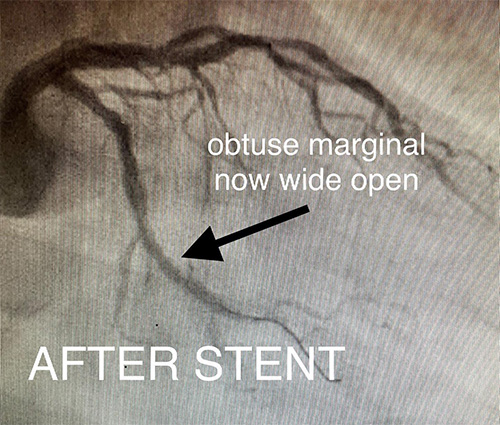
73 y/o female with Diabetes, found to have abnormal stress test prior to planned foot surgery. This lead to angiogram and subsequent percutaneous intervention of the ostium( origin) of the branch off the left circumflex artery called obtuse marginal. A single stent was used to open the blockage.
72 y/o female US National traveling from Mexico having chest pain.
57 y/o male with chest pain. He had an abnormal stress test which led to a CT angiogram imaging test showing a blockage. This led to the angiogram shown.
70 y/o female having “indigestion” which gets better with Sublingual nitroglycerin. Moreover, she had an abnormal stress test.
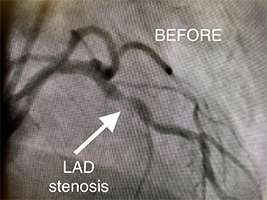
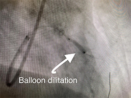
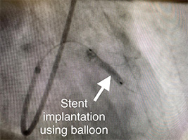
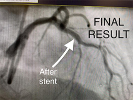
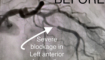
73 y/o female presents with accelerating symptoms of angina. Angiogram pictures show Left Anterior Descending artery (LAD) is severely blocked(stenosed). She receives an initial balloon dilatation and finally a drug eluting stent is implanted (see final result). She no longer has chest pain.
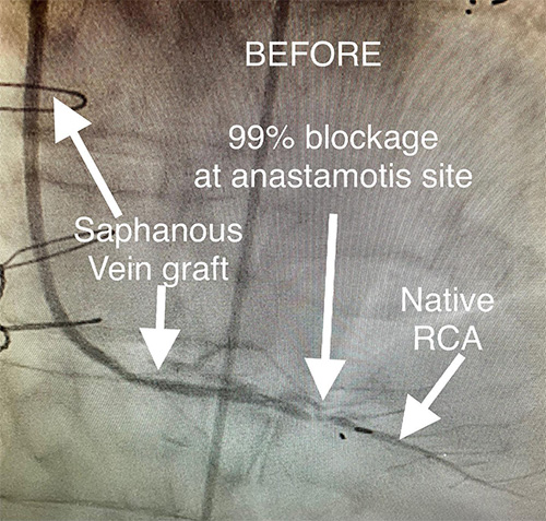
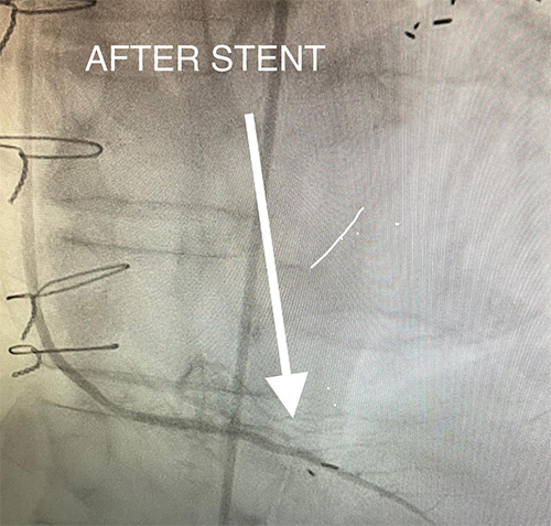
82 y/o man presents with new, worsening chest pain. He’s had a history of bypass surgery and multiple stents placed in the past. He was referred for coronary angiogram. Findings are seen in the “ Before” picture, followed by the therapeutic stent placed.
75 y/o female having chest pain with minimal exertion. After stent, no more chest pain.
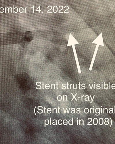
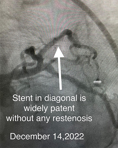
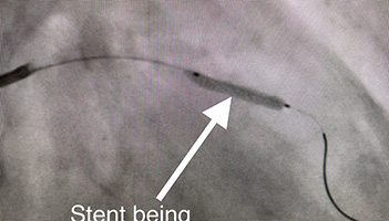
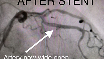
75 y/o female I placed a stent in her diagonal artery 2008, comes to my office complaining of shortness of breath and chest pain. First picture was X-ray showing silhouette of stent. Second picture shows coronary angiogram showing stent to be widely patent today December 14,2022 (14 years later).
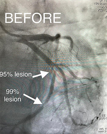
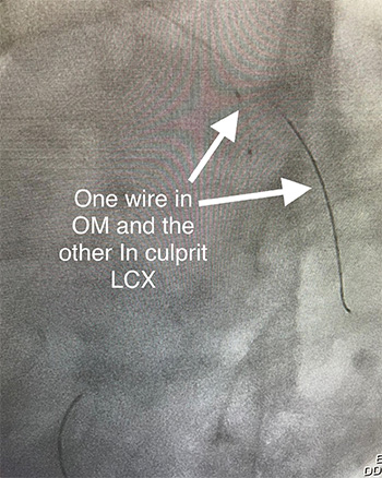
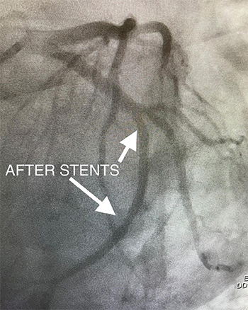
These are the 3 pictures of the 41 y/o male I described on earlier post. They show before (the blockages in the coronary arteries look like pinched off area), during and after the stents were successfully implanted. It’s imperative to remain on the anti-platelet medication for at least one year.
The coronary artery is what shows up in black. Contrast dye during X-ray appears black. The pinched off area in the “ before” picture is the stenosis. The “after” picture no longer shows the stenosis because a balloon opened it followed by a metal stent to prop it open. The balloon is removed, leaving only the metallic stent.
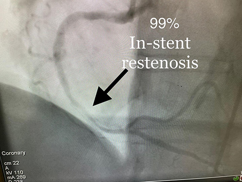
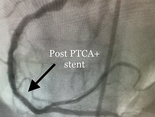
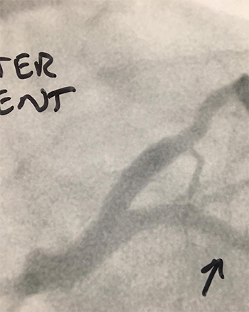
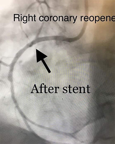
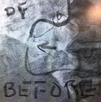
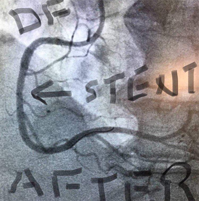
Women don’t get classic anginal chest pain. This is the perfect example: 82 y/o female who stated she had Right lower abdominal pain which is identical to her pain she had before her stent 10 years ago. After we reopened the stent restenosis, her abdominal pain resolved. She went home the day after her stent.
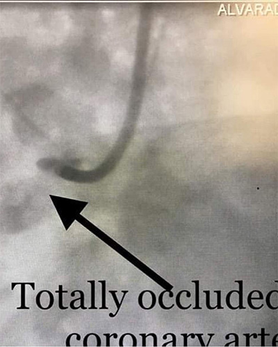
65 y/o male presents with acute chest pain and dizziness. EKG done in Emergency room shows ST elevation. Code STEMI called. The complete blockage was re-opened with a balloon and stent was placed. Symptoms and EKG improved. He went home 2 days later on anti-platelet medication he will need to stay on for one year.
An 58 y/o man presented with chest pain while traveling through San Diego. Before picture shows the tight blockage and “After” shows the results after emergency stent procedure performed by Dr. Nanavati.